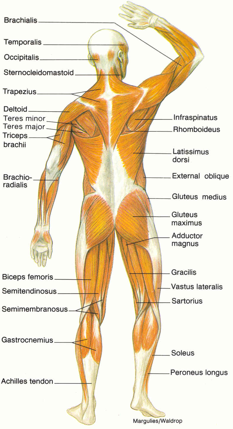
Labeled Muscles In The Body Diagram / Muscular System Rear (Labeled) - Body Part Chart Removable ... - You will also find extensor digitorum, extensor carpi group, latissimus dorsi, external oblique, gluteus medius, gluteus maximus, sartorius, peroneus longus, achilles tendon, gastrocnemius, hamstring group, flexor digitorum, triceps brachii, deltoid, trapezius, sternocleidomastoid, occipitalis in the.
Labeled Muscles In The Body Diagram / Muscular System Rear (Labeled) - Body Part Chart Removable ... - You will also find extensor digitorum, extensor carpi group, latissimus dorsi, external oblique, gluteus medius, gluteus maximus, sartorius, peroneus longus, achilles tendon, gastrocnemius, hamstring group, flexor digitorum, triceps brachii, deltoid, trapezius, sternocleidomastoid, occipitalis in the.. The free muscular system labeling sheet includes a blank diagram to label some of the main muscles in the body. The integumentary system consists of the skin, sweat and oil glands, nails, and hair. Skeletal, smooth and cardiac, according to the nih. Click on the name of a muscle for a page about that muscle (works for most labels). This quiz requires labeling, so it will test your knowledge on how to identify these muscles (latissimus dorsi, trapezius, deltoid, biceps brachii.
Label the muscles of the anterior forearm. There are around 650 skeletal muscles within the typical human body. Free online quiz label the muscle cell diagram. Use the location, shape and surrounding structures to. For instance, movement about the sagittal axis occurs in the sagittal plane e.g.
Posterior Muscles of the Human Body, illustration ... from media.sciencephoto.com The epidermis, dermis, and subcutaneous tissue. Studying these is an ideal first step before moving labeled diagram. Zygote body is a free online 3d anatomy atlas. Anatomical diagram showing a front view of muscles in the human body. This muscular system picture shows all the major muscle groups on the human body from the frontal view. Enchantedlearning.com label the body diagram label the human body diagram using the word list below. For instance, movement about the sagittal axis occurs in the sagittal plane e.g. You can click the image to magnify if you cannot see clearly.
An important group of muscles in the pelvis is the pelvic floor.
The pelvic floor muscles provide foundational support for the intestines and bladder. Skeletal, smooth and cardiac, according to the nih. The integumentary system consists of the skin, sweat and oil glands, nails, and hair. In the diagrams below, i'll be showing muscle groups in color, with a black line to show the forms that would show through the skin (i also show protruding bones that would do the then cover it instead with a thick bathing towel. Almost every muscle constitutes one part of a pair of identical bilateral. There are around 650 skeletal muscles within the typical human body. There is a printable worksheet available for download here so you can take the quiz with pen and paper. See how all sharpness disappears? The free muscular system labeling sheet includes a blank diagram to label some of the main muscles in the body. Label the muscles of the anterior forearm. Robins are cute, little songbirds in the united states. Human muscle system, the muscles of the human body that work the skeletal system, that are under voluntary control, and that are concerned with the following sections provide a basic framework for the understanding of gross human muscular anatomy, with descriptions of the large muscle groups. Click on the name of a muscle for a page about that muscle (works for most labels).
The integumentary system consists of the skin, sweat and oil glands, nails, and hair. Now label the diagram in your workbook! There are around 650 skeletal muscles within the typical human body. You can click the image to magnify if you cannot see clearly. Finally, the skeleton grows throughout childhood and provides a framework for the rest of the body to grow along with it.
Shoulder Muscles Diagrams | 101 Diagrams from www.101diagrams.com This is a table of skeletal muscles of the human anatomy. Muscle diagram anatomy practice health and physical education musculoskeletal system. Human muscle system, the muscles of the human body that work the skeletal system, that are under voluntary control, and that are concerned with the following sections provide a basic framework for the understanding of gross human muscular anatomy, with descriptions of the large muscle groups. The muscles labelled in the anterior muscles diagram shown above are listed in bold in the following table Skin is the largest organ in the body and is made up of three layers: The sartorius muscle is the longest muscle in the body. Enchantedlearning.com label the body diagram label the human body diagram using the word list below. Robins are cute, little songbirds in the united states.
Enchantedlearning.com label the body diagram label the human body diagram using the word list below.
The integumentary system consists of the skin, sweat and oil glands, nails, and hair. Muscles allow us to move and function. This is a table of skeletal muscles of the human anatomy. This image added by admin. Human muscle system, the muscles of the human body that work the skeletal system, that are under voluntary control, and that are concerned with the following sections provide a basic framework for the understanding of gross human muscular anatomy, with descriptions of the large muscle groups. Anatomynote.com found labelled diagram of the muscles in the human body from plenty of anatomical pictures on the internet. Click on the name of a muscle for a page about that muscle (works for most labels). Click on the labels below to find out more about your muscles. Rotation and hold ctrl down to pan the view. The soleus connects your lower leg bones to your heel, but it also gives your heart some help by pumping blood back. The muscular system can be broken down into three types of muscles: Almost every muscle constitutes one part of a pair of identical bilateral. Muscle diagrams are a great way to get an overview of all of the muscles within a body region.
Label the muscles of the anterior forearm. The free muscular system labeling sheet includes a blank diagram to label some of the main muscles in the body. There are around 650 skeletal muscles within the typical human body. Studying these is an ideal first step before moving labeled diagram. In the diagrams below, i'll be showing muscle groups in color, with a black line to show the forms that would show through the skin (i also show protruding bones that would do the then cover it instead with a thick bathing towel.
diagram of muscular system : Biological Science Picture ... from pulpbits.net Learn vocabulary, terms and more with flashcards, games and other study tools. Muscle diagrams are a great way to get an overview of all of the muscles within a body region. Use the location, shape and surrounding structures to. Skin is the largest organ in the body and is made up of three layers: Skeletal, smooth and cardiac, according to the nih. For instance, movement about the sagittal axis occurs in the sagittal plane e.g. Studying these is an ideal first step before moving labeled diagram. View, isolate, and learn human anatomy structures with zygote body.
Muscle diagrams are a great way to get an overview of all of the muscles within a body region.
View the muscles of the upper and lower extremity in the diagrams below. Click on the labels below to find out more about your muscles. There is a printable worksheet available for download here so you can take the quiz with pen and paper. When you are taking anatomy and physiology you will be required to identify major muscles in the human body. This is an online quiz called label the muscle cell diagram. Studying these is an ideal first step before moving labeled diagram. There are around 650 skeletal muscles within the typical human body. O skeletal hundreds of randomly located nuclei per cell (cells fuse together in development) striated. You will also find extensor digitorum, extensor carpi group, latissimus dorsi, external oblique, gluteus medius, gluteus maximus, sartorius, peroneus longus, achilles tendon, gastrocnemius, hamstring group, flexor digitorum, triceps brachii, deltoid, trapezius, sternocleidomastoid, occipitalis in the. View, isolate, and learn human anatomy structures with zygote body. The free muscular system labeling sheet includes a blank diagram to label some of the main muscles in the body. This muscular system picture shows all the major muscle groups on the human body from the frontal view. See how all sharpness disappears?
This image added by admin muscles in the body diagram. There is a printable worksheet available for download here so you can take the quiz with pen and paper.